
October 5, 2026: It was announced that Karl Deisseroth (Stanford University) would be awarded the Nobel Prize in Physiology or Medicine—an honor he shares with Peter Hegemann and Georg Nagel for their pioneering work on Optogenetics.
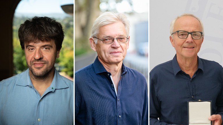
In 2018, Karl Deisseroth also received the Berthold Leibinger Future Prize for his work on “Lasers in the development and application of optogenetics.” He was nominated by Juergen Czarske, who had previously received the Leibinger Innovation Prize from TRUMPF (Ditzingen/Stuttgart) in 2008, together with Lars Buettner and Thorsten Pfister.
More under: Laser Preise
Other Nobel laureates among the recipients of the Leibinger Prizes include Stefan Hell (Leibinger Innovation Prize 2002, Nobel Prize in Chemistry 2014), Gérard Mourou (Leibinger Future Prize 2016, Nobel Prize in Physics 2018), and Anne L’Huillier (Leibinger Future Prize, September 2023, Nobel Prize in Physics, October 2023).
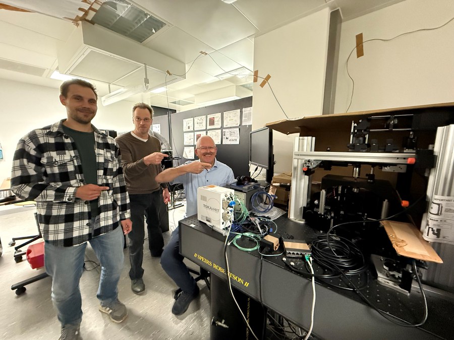
The Chair of Measurement and Sensor System Technology (MST) and the Competence Center Biomedical Computational Laser Systems (BIOLAS) have been conducting research on optogenetics for approximately ten years. A recent publication in Nature reported on a paradigm shift in optogenetics involving closed-loop measurement and control techniques:
R. Wendland, F. Schmieder, M. A. Sikandar, F. P. Knüppel, W.-H. Zimmermann, O. Bergmann, L. Büttner, J. W. Czarske, “All-optical closed-loop control of human cardiomyocyte networks exploiting holographic optogenetics,” Nature Communications Engineering 5, 159 (2026).
Dresden Researchers “Tickle” Cells with Light – Modeling Arrhythmias Using Holographic Optogenetics
The heart’s pumping function relies on the contraction of heart muscle cells (cardiomyocytes), which is triggered by electrical signals. The spatiotemporal excitation patterns generated by these signals are fundamental to synchronizing heart contractions and, consequently, maintaining normal cardiac function. Cardiac rhythm disorders (arrhythmias) primarily result from disturbances in the conduction of these excitation signals. For example, a spiral excitation wavefront can form, leading to an abnormally high heart rate (tachycardia) and potentially fatal ventricular fibrillation.
The mechanisms underlying the onset and termination of such disorders are not yet fully understood. These studies are conducted without animal testing and therefore support the 3R principles. The 3Rs—“replace, reduce, refine”—call for replacing animal experiments where possible, reducing their number to a minimum, and refining procedures to minimize the burden on animals.
In collaboration with the University Medical Center Göttingen, the team led by Dr. Lars Buettner and Prof. Juergen Czarske is modeling these phenomena using optogenetics—a technique that enables cellular activity to be controlled using light-sensitive proteins. The researchers use human cardiomyocytes derived from human induced pluripotent stem cells (iPSCs). By projecting specific spatiotemporal light patterns onto the cells, the researchers “tickle” them with light, thereby triggering defined excitation wavefronts.
Through in vitro experiments, they can specifically induce, observe, and control disturbances in excitation conduction. The aim of the current experiments was to detect sudden disturbances in the wavefront in real time and restore the normal state through adaptive light irradiation.
Image analysis and the calculation of computer-generated holograms for light-pattern generation—both essential components of the control loop—are computationally demanding because of the vast amounts of data involved. As a result, conventional approaches are unsuitable for real-time control. The researchers addressed this challenge by microscopically sampling the excitation wavefront at only selected points (“sparse sampling”). This drastically reduced the data volume and processing time while still allowing the state of the excitation wavefront to be identified unambiguously.
Using this approach, the researchers demonstrated, for the first time, the real-time control and restoration of a disturbed excitation wavefront.
For these experiments, the MST Chair operates the only certified Biosafety Level 1 (S1) laboratory within the Faculty of Electrical Engineering and Information Technology (ETIT) designed for work with genetically modified organisms (GMOs). The ability to conduct experiments directly on the cells in the laser laboratories was a crucial prerequisite for avoiding the stress that transportation could cause to the sensitive cell samples.
Through these experiments, the researchers demonstrated that optical systems engineering plays a key role in optogenetics as an enabling technology. They now intend to further develop their approach for applications involving more complex, three-dimensional cell structures. So-called organoids—physiologically relevant models—are considered to hold great potential across a wide range of fields, including disease modeling, drug screening, precision medicine, and regenerative medicine.
Nature publication:
R. Wendland, F. Schmieder, M. A. Sikandar, F. P. Knüppel, W.-H. Zimmermann, O. Bergmann, L. Büttner, J. W. Czarske, “All-optical closed-loop control of human cardiomyocyte networks exploiting holographic optogenetics,” Nature Communications Engineering 5, 159 (2026).
To the article on „All-optical closed-loop control of human cardiomyocyte networks exploiting holographic optogenetics“